The Physics Department
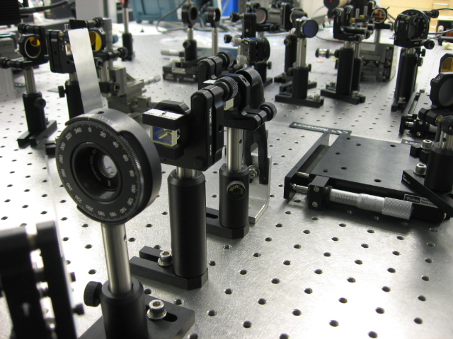
The optical coherence tomography (OCT) microscopy generates micron-scale three-dimensional images of living tissue down to depths of several mm. The OCT microscope, similar to a Michelson interferometer, detects depth information by interfering the backscattered beam from the sample with a reference beam. A full 3D image is constructed by varying the reference beam delay (axial - Z axis) and scanning the beam over the sample (X and Y axis). OCT has already became an important tool in ophthalmology but technical challenges limit its use in other applications.
We approach the problem of medical imaging from the ultrafast optics perspective. If we can completely characterize an optical pulse that backscatters from a sample, we can deduce a great deal of information about the sample. By integrating optical coherent tomography into a laser micromachining platform, we are able to follow the extreme sample morphology changes in real-time at rates up to 312 kHz. This novel diagnostic provides a new perspective into the extreme light-matter interaction. It's also a lot of fun and has been making waves in industry!