
Science unlocks the mysteries of the world through objective data and numbers. But what if researchers could explain their work through art? Queen’s put that question to its research community in its annual Art of Research photo and video contest, and Queen’s researchers responded with a stunning array of images and videos of groundbreaking work taking place in and out of the lab.
Now in its eighth year, the contest invites faculty, staff, students, and alumni to reveal the beauty behind discovery. Images from the competition take the public behind the scenes, offering a rare glimpse into the people, places, and moments at the heart of research.
The categories this year were inspired by Queen’s Strategic Research Plan 2025–2030 and the university’s Bicentennial Vision, reflecting priority research areas such as health and well-being, next-generation computing, sustainable societies, materials innovation, and our understanding of the Earth and universe.
These photographs tell stories of researchers working at the forefront of some of the world’s most urgent challenges – advancing medical discoveries, exploring artificial intelligence, and addressing climate change, social equity, and the future of sustainable communities. The contest also highlights the strength of the Queen’s research ecosystem, where scholars are supported with the expertise, facilities, and environment needed to pursue ambitious ideas. As outlined in the university’s Bicentennial Vision, Queen’s is sharpening its focus on areas where it can lead nationally and globally, ensuring its research continues to deliver meaningful impact in Canada and around the world.
This photo essay showcases this year’s photography winners, giving researchers a chance to explain their work in their own words.
-
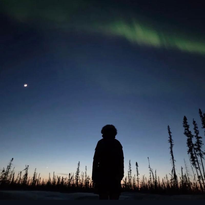
Photograph by Lydia Morrow

Category: Building Inclusive and Sustainable Societies
Title: Still Standing
Researcher: Lydia Morrow, MASc student, civil engineering
Queen’s Lab: Cryo-Hydro(geo)logy Interdisciplinary Laboratory (CHIL)
Location: Scotty Creek Research Station, on the lands of the Łı́ı́dlı̨ı̨ Kų́ę́ First Nation, Northwest Territories
A researcher pauses under the northern lights at Scotty Creek Research Station on Łı́ı́dlı̨ı̨ Kų́ę́ First Nation territory, surrounded by the ghostly silhouette of trees killed by a 2022 wildfire. That event sits at the heart of my research. My work examines how wildfire and permafrost thaw affect mercury cycling and water quality in subarctic peatlands. These landscapes are growing increasingly vulnerable as wildfire frequency and intensity continue to rise across the Canadian subarctic.
-
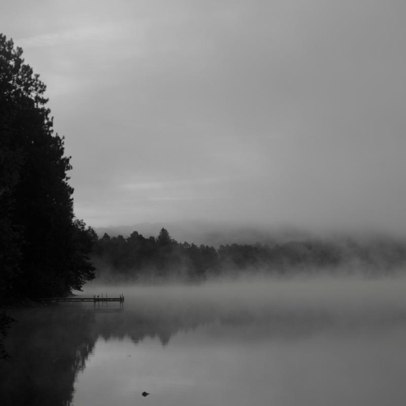
Photograph by Dakota Gustafson

Category: Special Prize – Narratives of Discovery
Title: Haze
Researcher: Dakota Gustafson, MD–MSc Student, public health sciences and medicine
Queen’s Lab: Brooks Lab
Location: Barry’s Bay, Ont.
The mind after cardiac arrest does not return unscathed. It arrives the way morning comes to still water: slowly, incompletely, shadowed by something the doctors didn’t warn you about. Haze captures that threshold state. The treeline exists, the dock is there, yet everything is heavy, muted, wrong in ways that are hard to name. Your life is reflected back at you, but you can no longer feel it the way you once did. Depression, anxiety, PTSD – these are the fog that settles after the heart restarts. My research asks the first necessary question: how many people are living inside it?
-
Photograph by Axelle Chevalier-Héroux

Category: Envisioning Just Futures
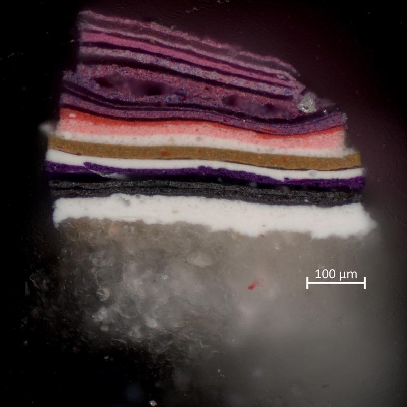
Title: Free Fall – Paint Cross-Section
Researcher: Axelle Chevalier-Héroux – MAC student, art conservation
Location: Art conservation, Queen’s University
This photo, taken with an optical microscope, depicts a cross-section of the layers of acrylic paint on a sample of Free Fall (circa 1973), an op-art installation made by the Canadian artist Frank Barry (1913–2013). It was captured during material analysis to explore the layered structure beneath the surface of the panel painting. The cross-section reveals a complex colour stratigraphy, with up to 20 layers and colour variations – such as white, grey, purple, ochre, bright pink, and red – that would not otherwise be visible. It highlights the artist’s understanding of colour theory and a trial-and-error process.
-
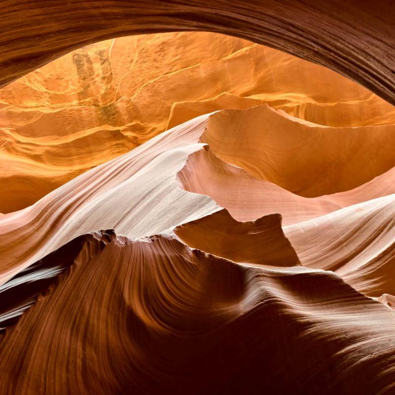
Photograph by Tianyu Zhao

Category: Understanding the Earth and Universe
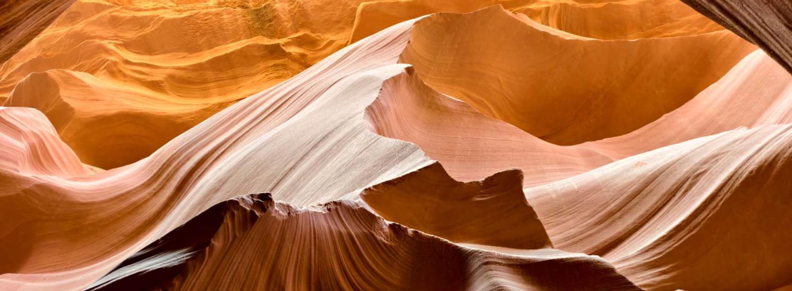
Title: Earth’s Layered Legacy: From Rock to Resource
Researcher: Tianyu Zhao, post-doctoral fellow, Robert M. Buchan Department of Mining
Queen’s Lab: Hydrometallurgy and Environmental Group
Location: Antelope Canyon, Ariz.
These layered sandstone waves of Antelope Canyon are shaped by millennia of water and wind. They mirror the geological strata we study to locate and extract mineral resources. As a mineral scientist, my work builds on these natural processes to responsibly explore and utilize Earth’s mineral wealth. This image is a reminder that our most valuable resources are written in the rocks around us, a timeless testament to the planet’s quiet generosity and our shared duty to steward it wisely.
-
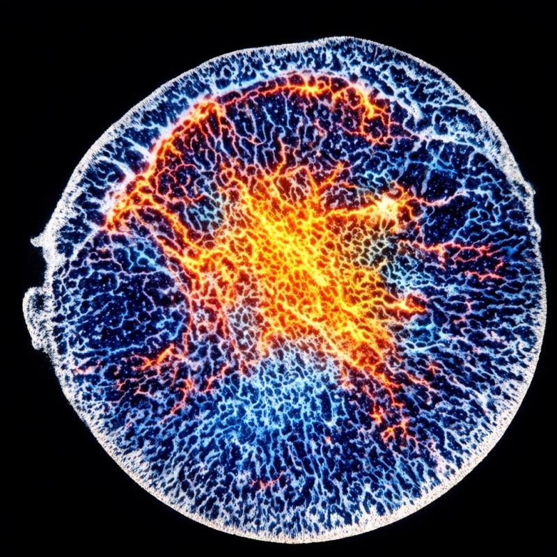
Photograph by Emily Cameron
Category: Special Prize – Narratives of Discovery
Title: Stories Written in Our Bones
Researcher: Emily Cameron – PhD student, mechanical and materials engineering
Queen’s Lab: Queen’s Small-Animal Imaging Laboratory (SAIL) and Centre for Health Innovation
Location: Queen’s University and Kingston Health Sciences Centre
At first glance, bone appears solid and unchanging. However, it is constantly adapting to how we move. This image shows the inside of a femoral head, the ball of the hip joint that carries us through life. Over years of walking, working, and playing, bone strengthens along the pathways used most often. These changes build an intricate internal architecture shaped by a person’s life in motion. These pathways resemble lightning across the sky, roots spreading through soil, or tree rings quietly recording a life lived. My research reads these patterns to design implants that work with the body’s natural architecture.
-
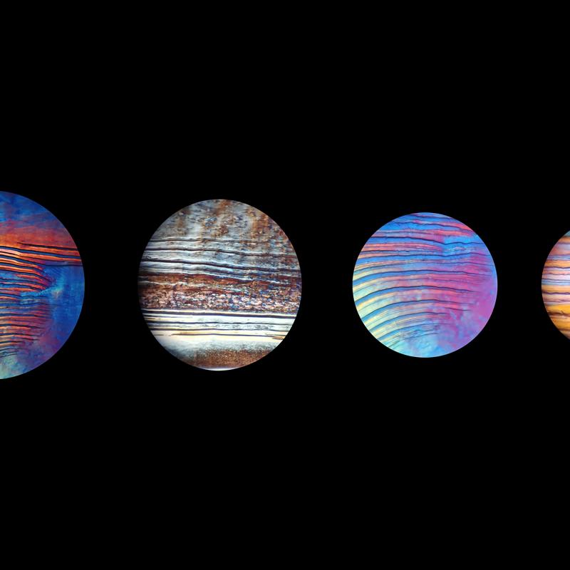
Photograph by Mostafa Rammal

Category: Delivering Materials for the Future
Title: Planets from Plants
Researcher: Mostafa Rammal, post-doctoral fellow, chemical engineering
Queen’s Lab: De France Lab
Location: Biosciences Complex, Queen’s University
What appears to be from a galaxy far, far away is actually the self-assembly of plant-derived cellulose nanocrystals during evaporation, captured using a polarized light microscope. As water evaporates, capillary flow drives the suspended nanocrystals toward the edge, forming the well-known coffee-ring effect. During this process, the rod-like cellulose nanocrystals spontaneously organize into twisted crystalline structures. These ordered regions produce iridescent colours that resemble astronomical structures. Understanding these assemblies is essential to my research, which aims to integrate these nanocrystals with polymers to engineer high-performance, sustainable materials.

