 Research at Queen's
University Animal Care Committee Standard Operating Procedure
Document No: 7.11
Subject: Retro-orbital Injections in Mice
Date Issued: April 11, 2012
Revision: 3
Location: Queen’s University
Responsibility: Principal Investigators (PI), Research Staff, Veterinary Staff
Purpose: The purpose of this Standard Operating Procedure (SOP) is to describe the standard method used for retro-orbital injections in mice.
1. Introduction and Definitions:
Retro-orbital injections are commonly used for transplants of stem cells (not limited to) as an alternative to tail vein intravenous injections. They are less challenging technically than tail vein injections. Because the needle is being placed in the retro-bulbar space (the region behind the globe of the eye) the mouse should be anesthetized so that it remains still during the procedure.
Abbreviations:
Animal Care Services ACS, Principal Investigator PI, subcutaneous SC, intravenous IV, intraperitoneal IP, intramuscular IM, per os PO, per rectum PR
2. Materials:
- 1 cc sterile syringe (or insulin syringes)
- 27-30g sterile needles
- Anesthesic (isoflurane vaporizer, injectable anesthetic)
- Induction chamber and nose cone
- Sterile gauze
- Ophthalmic anesthetic (i.e. 0.5% proparacaine hydrochloride ophthalmic solution - kept in fridge)
- Absorbent pad
3. Procedures:
- Each and every animal requires a new sterile syringe and a new sterile needle.
- Load the sterile 1cc syringe and a 27-30g needle with appropriate volume to be injected (maximum 200uL).
- It is imperative that the material being injected contains no clump material (always filter cell suspension prior to injection).
- Instill a drop of ophthalmic anesthetic.
- The procedure is facilitated by being seated at a table bench or hood.
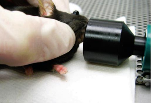
- Anaesthetise the mouse (following SOP 7.6 Anesthesia in Mice). The animal must be maintained under anesthesia via a nose cone until the handler is proficient in the procedure.
- When the mouse has lost its righting and pedal reflex, with your non-dominant hand, scruff it securely by the nape of the neck, with tail wrapped around little finger (SOP 7.20 Manual Restraint of Mice).
- Place the mouse on the absorbent pad on the table surface and transfer to nosecone.
- Skilled personnel who do not require extended anesthesia can cup the scruffed animal in their palm if preferred.
- If the scruff is insufficient to protrude the globe, gently retract the fur above the eye. The eye must bulge slightly.
- Position needle tip on the medial canthus and apply mild downward pressure, with the bevel facing downwards.
- Contrary to most injections, this position (bevel away from globe and towards the medial canthus - refer to image) reduces the possibility of damage to the globe. The needle should be maintained at a 30-45 degree angle relative to the medial canthus. Carefully advance the tip of the needle to penetrate the retro-orbital sinus.
- The needle often touches an orbital bone, providing user with a distinct landmark. Stop advancing at this point (approximately mid sinus). It is helpful to withdraw the needle 1mm at this point to ease the injection flow.
- Do not aspirate.
- Inject up to 150uL for adult mice and up to 10 uL for infant mice slowly, examining the globe closely. The user must stop immediately if swelling or bleeding is observed. There should be no resistance during infusion.
- After the injection, the needle should be slowly and smoothly withdrawn. Pivoting the needle slightly before removal breaks the last bead of compound and helps to prevent leakage.
- Following recovery from anaesthesia, the animal should be observed until they are conscience and ambulatory and then returned to their cage.
- For multiple injections at least 48 hours should elapse between injections and the eyes should be alternated.
- Although retro-orbital injections are less traumatic than retro-orbital blood sampling, eye trauma still can occur and the eye should be inspected post-procedure, and 1-day post-procedure.
- In the event the eye is damaged and/or becomes opaque, injections must stop and the veterinarian must be consulted for treatment.
- No injections may be performed in a damaged eye.
.png)
(Illustration by Darryl Leja)
Correct placement of the needle relative to the retro-orbital sinus, the globe and the back of the orbit.
The mouse’s eye is partially protruded from the socket by applying gentle downward pressure to the skin dorsal and ventral to the eye.
Yardeni T, Eckhaus M, Morris HD, Huizing M, Hoogstraten-Miller S. Retro-orbital injections in mice. Lab Anim (NY). 2011 May;40(5):155-60.
| Date | New Version |
|---|---|
| 02/28/2019 | Triennial Review |
| 02/28/2022 | Triennial Review |
| 01/22/2025 | Triennial Review - Updated format and wording |